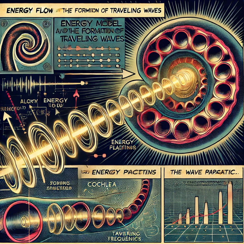
Simplified Model to Demonstrate the Energy Flow and Formation of Traveling Waves similar to those found in Cochlea
The hearing nerves of vertebrates have their endings on the basilar membrane. The total length of this membrane in man is 35 mm. Its width varies continuously from 0.04 to 0.5 mm., and accordingly the stiffness of the membrane decreases over its length one hundred fold. The whole membrane is imbedded in fluid, and, when it is set in vibration, waves travel from the stiff part of the membrane toward the softer part. The properties of these waves are of major interest.
http://www.pnas.org/content/42/12/930.full.pdf+html
To create a simplified model demonstrating the energy flow and formation of traveling waves similar to those found in the cochlea, we can use a simple mechanical system that mimics the basic principles involved. Here’s a basic conceptual model:
1. Mechanical Oscillator: Start with a basic mechanical oscillator, such as a string or a thin elastic membrane, stretched between two fixed points. This oscillator represents the basilar membrane in the cochlea.
2. Excitation Source: Apply a periodic excitation to the oscillator to simulate sound input. This excitation could be in the form of a periodic force or displacement applied to one end of the oscillator.
3. Propagation of Waves: When the excitation is applied, waves will propagate along the oscillator, similar to how sound waves travel along the basilar membrane in response to sound stimuli. The waves will travel from the point of excitation toward the opposite end of the oscillator.
4. Frequency-Dependent Response: The oscillator should exhibit frequency-dependent response, meaning that different parts of the oscillator will resonate or vibrate preferentially at different frequencies. This mimics the tonotopic organization of the cochlea, where different regions respond maximally to different frequencies.
5. Traveling Waves: As the waves propagate along the oscillator, they will induce traveling waves, causing localized displacement or vibration of the oscillator at specific points along its length. These traveling waves represent the spatial distribution of energy along the basilar membrane in response to different frequencies.
6. Detection and Analysis: Finally, incorporate a method for detecting and analyzing the displacement or vibration patterns of the oscillator. This could involve using sensors or transducers placed at different points along the oscillator to measure the magnitude and phase of the vibrations.
By observing the displacement or vibration patterns of the oscillator in response to different frequencies of excitation, you can demonstrate the formation of traveling waves and the frequency-dependent response similar to those observed in the cochlea. This simplified model provides a visual and tangible representation of the basic principles involved in cochlear mechanics and the generation of auditory signals.